AmnioBind
Regenerative medicine plays an important role in the care continuum. Using human cell and tissue products to repair, restore and replace damaged tissue gives patients and professionals options that weren’t available just a few years ago.
Introducing AmnioBind™
AmnioBind™ contains growth factors cytokines, glycoconjugates, glycosaminoglycans and a comprehensive collagen matrix. These naturally occurring factors in the placental membrane are preserved to provide AmnioBind™ the necessary elements required to function as a biological covering.

What is AmnioBind?
Biologically active wound cover
AmnioBind™ is a dehydrated intact placental membrane covering that preserves the naturally occurring cytokines, growth factors, comprehensive collagen matrix, glycoconjugates, and glycosaminoglycans.
Our in-house propriety process allows preservation of the placental membrane with little to no degradation of the naturally occurring proteins during the dehydration and sterilization process. Upon the application of AmnioBind™, the cytokines, growth factors, as well as the structural proteins are returned to a near-native state which provides an ideal protective barrier for wounds.
By preserving all layers of the placental membrane, AmnioBind™ is a protective barrier with exceptional structural integrity.
Why AmnioBind?
Protecting biology
Similar products are processed utilizing excessive scraping, membrane separation, cross linking, chemicals, and high temperatures which reduce the levels of available growth factors and cytokines and can affect the structural integrity of the membrane.
We do not separate the amnion and chorion layers and re-combine the tissue. AmnioBind™ is an intact placental membrane that utilizes a propriertary processing method which uses no unnecessary chemicals, antibiotics, or preservatives, and is never frozen.
By preserving the natural state of the placental membrane we are able to provide an ideal protective barrier for wounds that maintains the functional proteins found in the tissue.


Placental Membrane
A Mother’s Gift
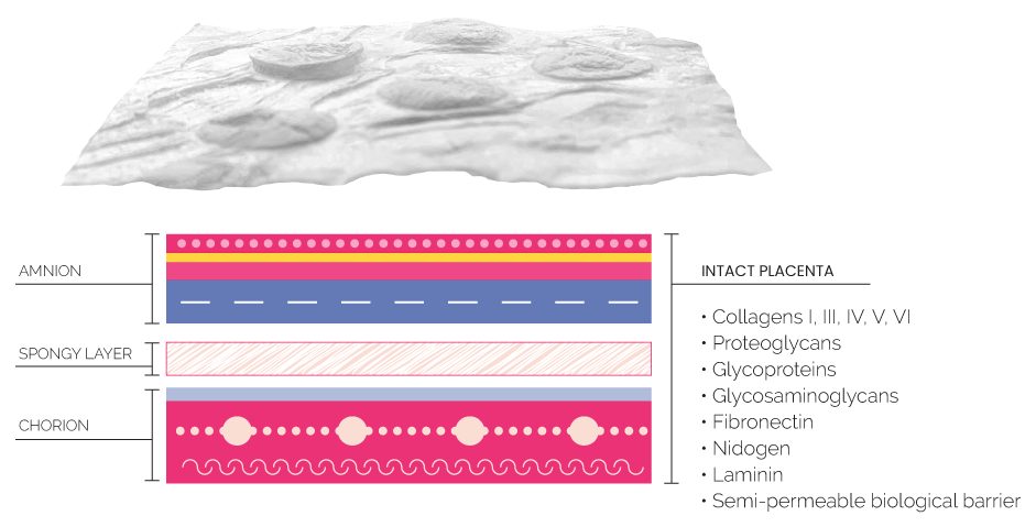
The placental membrane is a biological structure devoid of nerves, muscles or lymph vessels. It receives nutrients and oxygen from the chorionic uid and transfers it across the membrane to the fetal surface vessels.1 As it protects the growing fetus from the pressures of surrounding structures, it also facilitates metabolic functions, such as the transport of water and soluble materials, and produces important biological factors, such as cytokines and growth factors.2
The fetal membranes consist of two layers: the outer chorion which is in contact with the uterine wall, and the amnion (amniotic membrane) which lies on the fetal side and is in contact with amniotic uid. The amniotic membrane’s basal surface lies on top of the chorion, the outer most layer of the placental membrane.3 The amniotic membrane and the chorion are not bonded and remain separable even after delivery.4
The Makeup Of Birthing Tissue
What is found in birthing tissue that can be useful for wound covers? Aside from having live cells, the most abundant components by mass within a tissue like the umbilical cord is collagen, primarily Collagen Type I. These birthing tissues also contain other common molecules of the Extracellular Matrix (ECM) including more structural proteins like fibronectin and fibrillin, smaller proteins like growth factors and cytokines, and glycosaminoglycans such as hyaluronic acid(1–3).
These tissues contain a wide variety of growth factors and cytokines, with roughly 500 different soluble proteins identified so far4. Including transforming growth factor-beta 1 (TGF-β1), platelet derived growth factor-AA (PDFG-AA), vascular endothelial growth factor (VEGF), and angiogenin-4, all known to be involved in cell proliferation(3).
Why These Components Matter | Factors Found in Placental Membrane
Collagens are the most abundant proteins in the body, yet many studies show that readily available collagen added into a wound space contributes to wound healing, aid in the formation of blood vessels and arteries, and can reduce time to heal(5–7). Studies like these are the basis for the large influx of collagen dressings.
Hyaluronic acid has also been shown on multiple occasions to aid in the healing of wounds(8–11). Not only does birthing tissue contain a high concentration of hyaluronic acid, but when used as a wound dressing it has the potential to form into a natural hydrogel dressing, depending on how the tissue is processed.
These hyaluronic acid-based gels are exceptional at retaining moisture and can absorb much of the fluid which exudes from many wounds. Although not a pretty picture, these performance characteristics are vital when healing difficult wounds such as ulcers. Furthermore, the soluble proteins fulfill a host of important roles in wound healing. In addition to cell proliferation described previously, these cytokines and growth factors modulate inflammation, regulate cell growth, aid in revascularization, among many others(3).
WORKS CITED
1. Restb M.Moradi; Améli, S. FrancaJ. ; C. R. R. G. M. van der. Microfibrillar composition of umbilical cord matrix: Characterization of fibrillin, collagen VI and intact collagen V. Placenta (1998).
2. Azusa Matsumotoa; Terue Kawabataa; Yasuo Kagawaa; Kumiko Shojia; Fumiko, Kimurabc; Teruo, Miyazawad; Nozomi, Tatsutae; Takahiro, Arimaf; Nobuo, Yaegashig; Kunihiko, N. Associations of umbilical cord fatty acid profiles and desaturase enzyme indices with birth weight for gestational age in Japanese infants.
3. Barrientos, S., Stojadinovic, O., Golinko, M. S., Brem, H. & Tomic-Canic, M. PERSPECTIVE ARTICLE: Growth factors and cytokines in wound healing. Wound Repair and Regeneration 16, (2008).
4. Bullard, J. D. et al. Evaluation of dehydrated human umbilical cord biological properties for wound care and soft tissue healing. Journal of Biomedical Materials Research Part B: Applied Biomaterials 107, 1035–1046 (2019).
5. Fleck, Cynthia A.; Chakravarthy, D. Understanding the Mechanisms of Collagen Dressings. Advances in skin & wound care (2007).
6. Karr, J. C. et al. A morphological and biochemical analysis comparative study of the collagen products Biopad, Promogram, Puracol, and Colactive. Advances in skin & wound care 24, 208–216 (2011).
7. Schultz, G. S. & Wysocki, A. Interactions between extracellular matrix and growth factors in wound healing. Wound Repair and Regeneration 17, 153–162 (2009).
8. M Prosdocimi 1, C. B. Exogenous hyaluronic acid and wound healing: an updated vision. Panminerva Med. (2012).
9. Nyman, Erika MD, PhD; Henricson, Joakim PhD; Ghafouri, Bijar PhD; Anderson, Chris D. MD, PhD; Kratz, Gunnar MD, P. Hyaluronic Acid Accelerates Re-epithelialization and Alters Protein Expression in a Human Wound Model. Plastic and Reconstructive Surgery (2019).
10. Véronique Voinchet 1, Pascal Vasseur, J. K. Efficacy and safety of hyaluronic acid in the management of acute wounds. American Journal of Clinical Dermatology (2006).
11. Litwiniuk, M., Krejner, A. & Grzela, T. Hyaluronic Acid in Inflammation and Tissue Regeneration. Wounds (2016).
© 2022 NuWave Med Healthcare Solutions | Brandana Marketing Solutions

